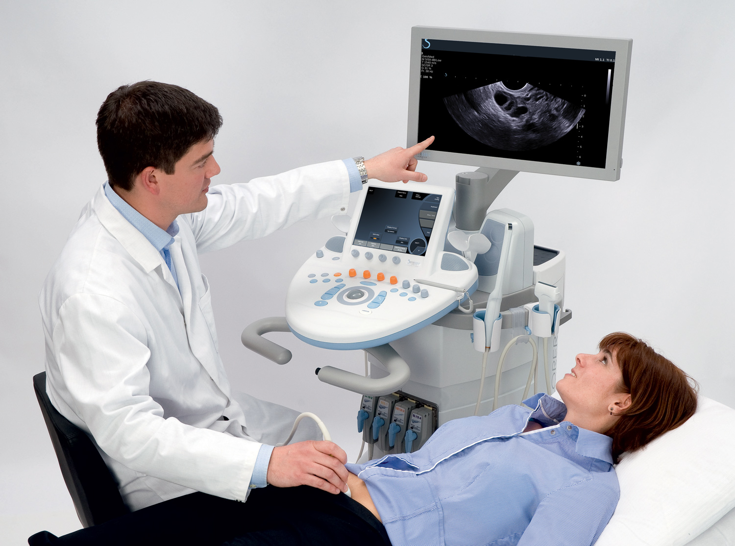
GYNAECOLOGY
|
|

|

|
|
Clarity Matters
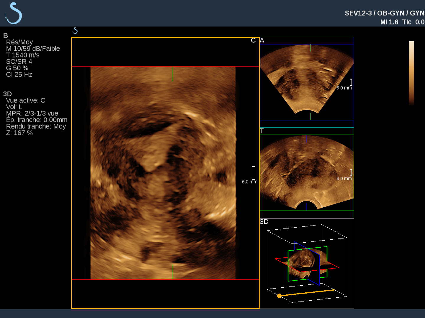
The advanced visualization capabilities of SuperSonic™ MACH™ systems let you clearly see fine morphological structural details of the ovaries, adnexae and endometrium, including difficult cases such as a fibroid uterus.1,2
Optimized factory pre-sets make it easy to quickly obtain the clearest gynecologic images possible, saving you exam time, while ultrasensitive Doppler capabilities aid you in identifying vascular abnormalities of the uterus.
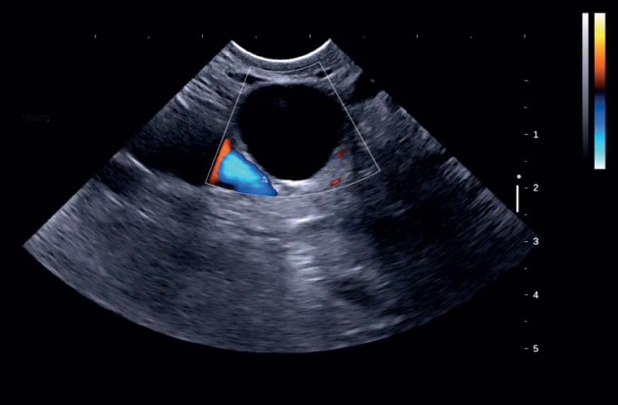
Additional tools such as SuperCompound™ ensure your image quality is not compromised while in Color Flow.
Let ShearWave Elastography™ (SWE), without manual compression, improve your diagnostic confidence through real-time, quantitative, color-coded tissue elasticity visualization. SWE could be especially helpful in determining new growth of fibroids, confirming cystic, complex or solid ovarian masses, and assessing the follow-up of post-fibroid embolization.
Transducers

| SuperEndocavity™ SE12-3
Number of elements: 192 | |

| SuperEndocavity™ Volumetric 12-3
Number of elements: 192 | |

| Single Crystal Curved XC6-1
Number of elements: 192 |
References:
1 Engineering Clinical Evaluation (Ece) V10 Endocavity Probes Evaluation in Gynecology Dr Shojai Aix En Provence; PM.TP/TR.034
2 V10 CMR Validation – Institut de Radiologie de Paris – Gynecology; PM.TP/TR.036

