LIVER
|
|

|
|
|

|
|
See What You Measure
Liver markers in UltraFast Ultrasound Imaging
SuperSonic Imagine innovates by introducing new markers which allow images to be associated with quantitative values. These measurements of fat content and tissue viscosity may help better manage patients with chronic liver disease.
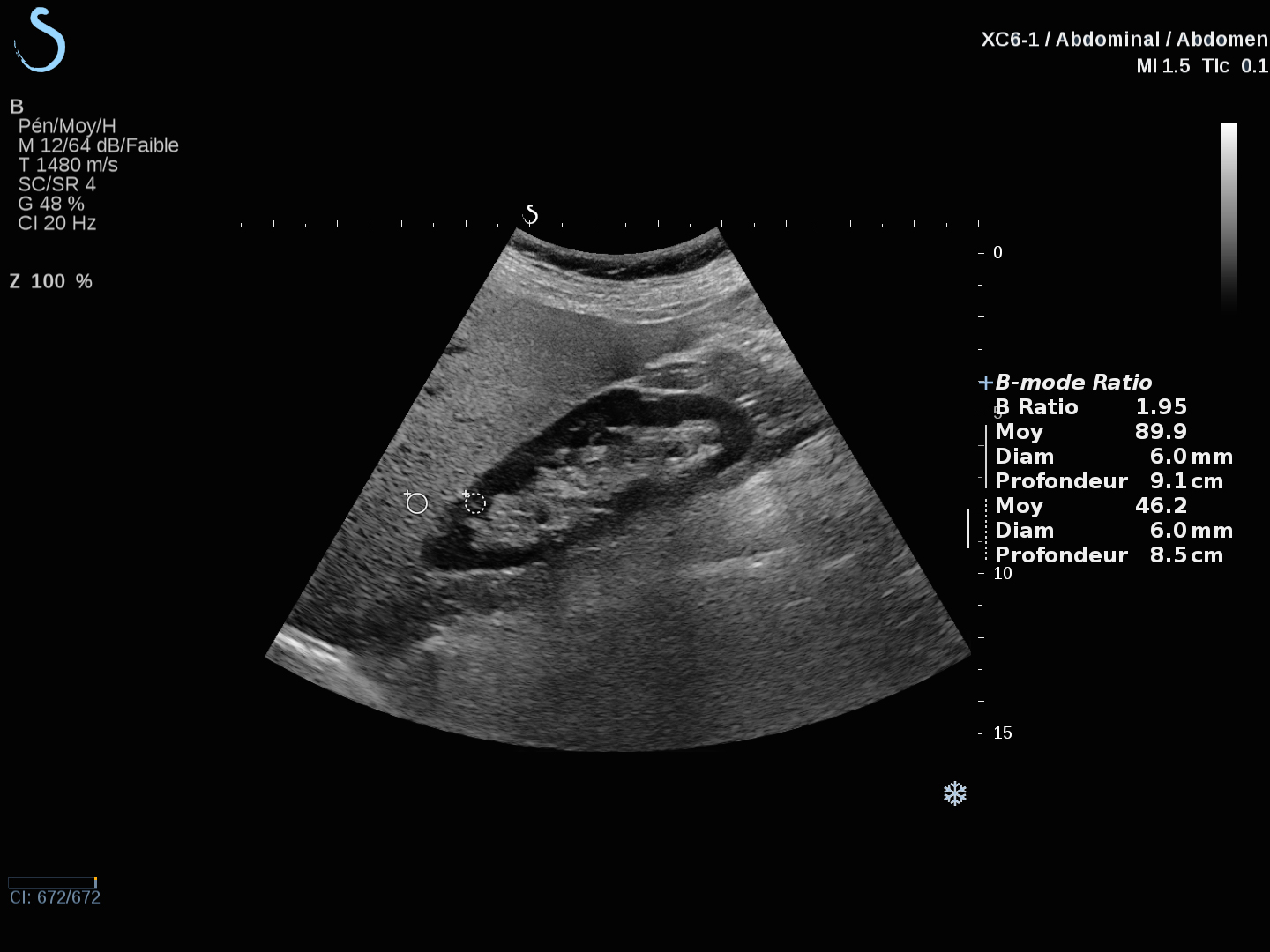
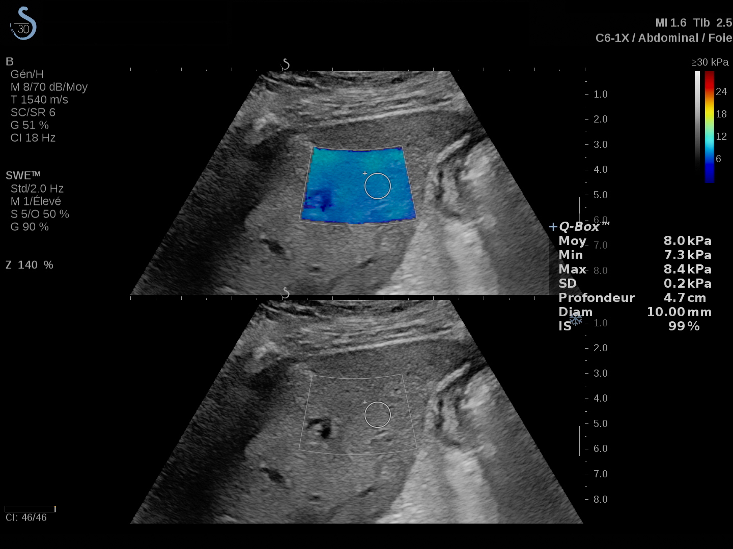
Real-time ShearWave™ Elastography
ShearWave Elastography (SWE™) is a quick, non-invasive, reporducible exam that provides quantitative measurement and color-coded maps of the tissue stiffness on an anatomic B-Mode image. It can be performed on patients with ascites.
- Can be performed in a hospital or private practice
- Takes as little as 60 seconds
- Provides imaging and liver stiffness quantification simultaneously
More than 160 publications have demonstrated the reliability and effectiveness of SWE for the assessment of liver fibrosis in patients with chronic hepatitis B, C and NALFD.
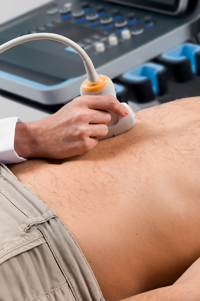
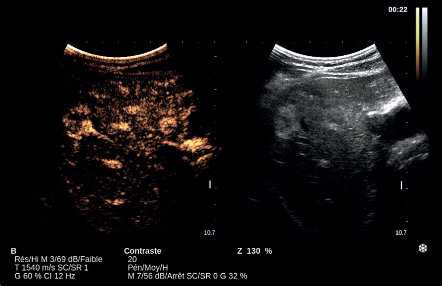
B-Mode ratio
Hepato-renal contrast calculation can evaluate the level of steatosis;
Transducers

| Single Crystal Curved Array XC6-1
Number of elements: 192 |
References:
1 Fujiwara et al., The B-mode image-guided ultrasound attenuation parameter accurately detects hepatic steatosis in chronic liver disease, Ultrasound in Med. & Biol. 2018
2 Dioguardi Burgio et al., Ultrasonic Adaptive Sound Speed Estimation for the Diagnosis and Quantification of Hepatic Steatosis: A Pilot Study, Ultraschal Med. 2018
3 Deffieux T et al., Shear Wave Spectroscopy for In Vivo Quantification of Human Soft Tissues Visco-Elasticity, IEEE Transactions on Medical Imaging, 2009

