Supersonic® MACH® Key Features

Explore our advanced features that enhance your diagnostic capabilities and improve user experience.

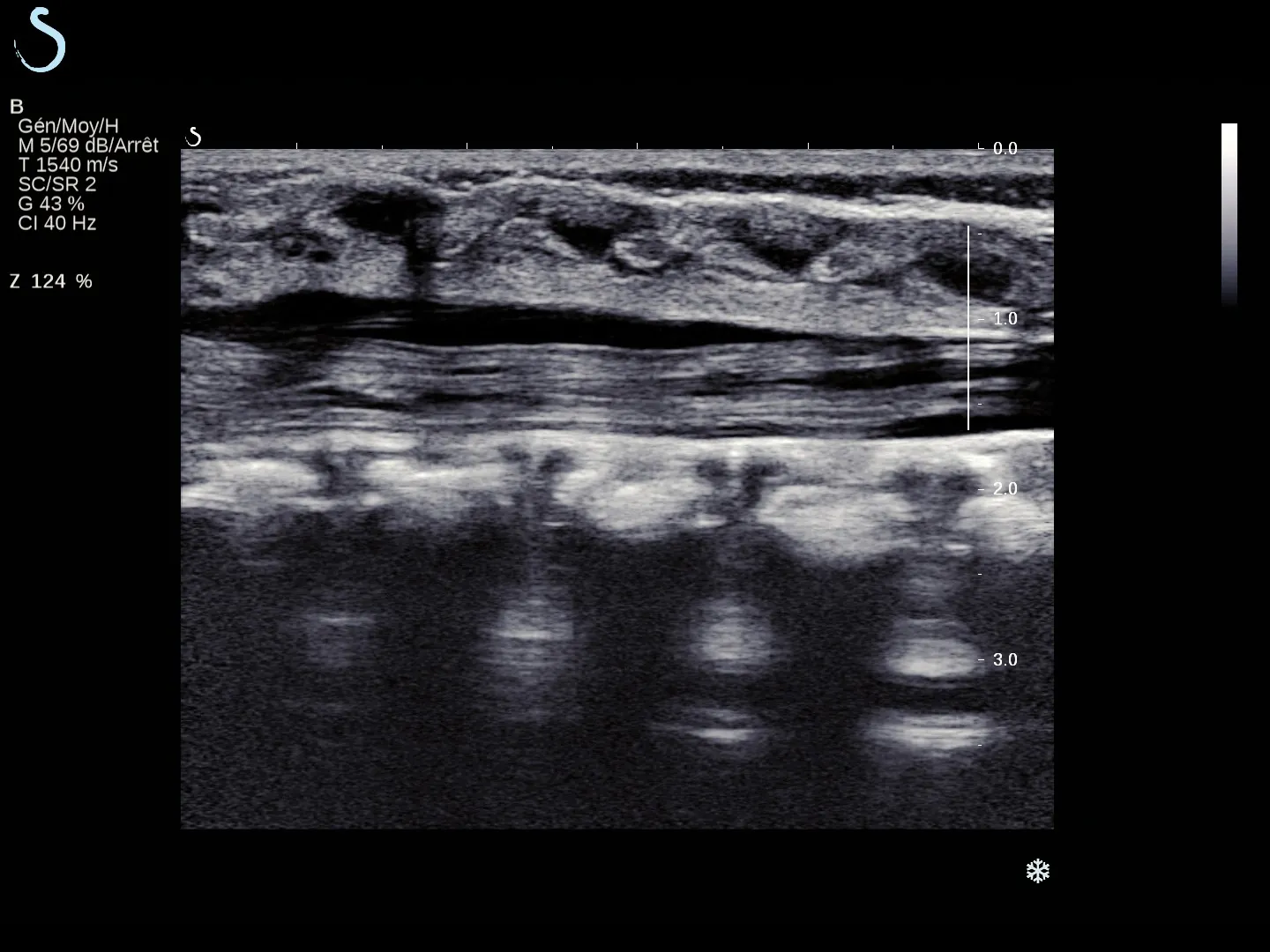
Unparalleled Image Quality

Unparalleled Image Quality

The innovative transducer design and powerful capabilities of software-based architecture are optimizing signal-to-noise ratio at each step of signal processing, offering you an incredible definition in both fundamental and harmonic imaging modes.(2, 3, 4)

A set of advanced features is available to simplify and speed up the image acquisition process.

These include:

  • Optimized penetration settings: for visualizing structures at variable depths and in dense breast
  • SuperCompound™: fast compounding designed for smooth images with reduced speckle
  • SuperRes™: delineation of structures to help improve lesion conspicuity
  • TissueTuner™: clear images obtained by matching the speed of sound to tissue density
  • AutoTGC™: designed for optimization of the gain for the entire image
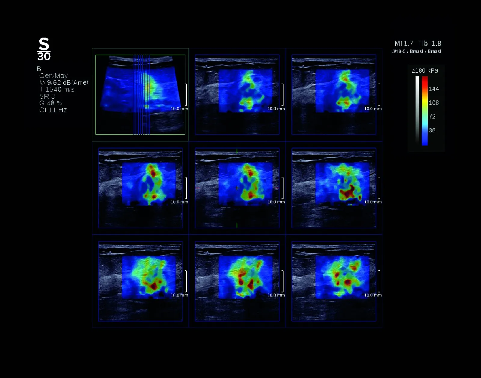
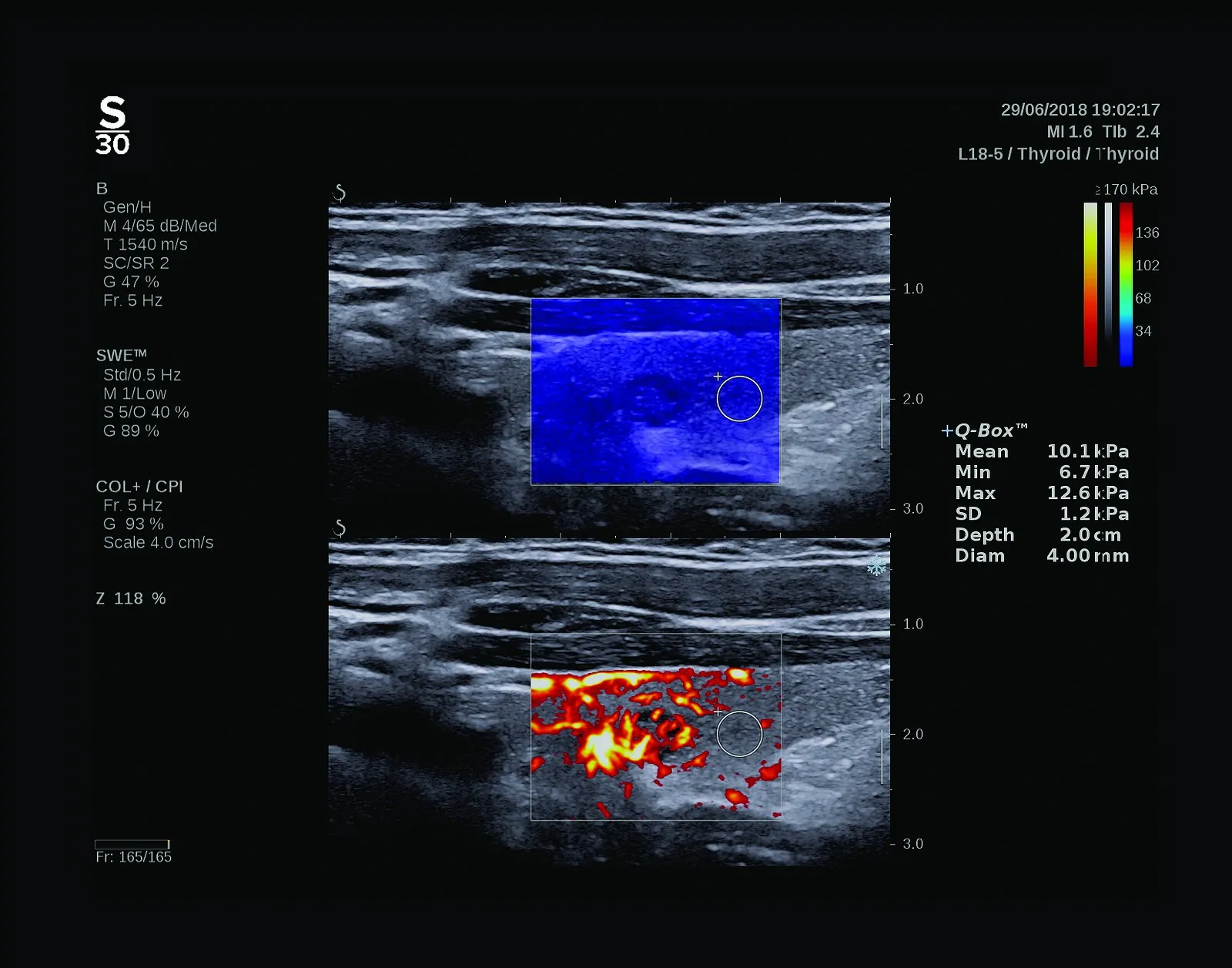
Real-Time ShearWave® PL.U.S.™ & 3D ShearWave®

Real-Time ShearWave® PL.U.S.™ & 3D ShearWave®

Real-Time ShearWave® Elastography

  • ShearWave Elastography Pioneers, first to introduce ShearWave® in real time in 2008
  • Superior dynamic non-invasive tissue stiffness evaluation
  • ShearWave® comes standard on all transducers*
  • Largest ROI on the market
  • Available in 2D and 3D imaging
  • No lag time, no compromise to b-mode image quality
  • 1000+ peer-reviewed publications

*Disabled on the P5-1X transducer

Real-Time 3D ShearWave® Elastography

  • Aids in the diagnostic workup of breast lesions and positively impacts patient management (5)
  • Contributes to more accurate tumor size estimation and clear margin definition in the preoperative setting (6)
  • Plays a role in monitoring and evaluation of breast cancer patients during and after neoadjuvant chemotherapy (7)
  • 5. Chen YL, Chang C, Zeng W, et al. 3 Dimensional shear wave elastography of breast lesions: Added value of color patterns with emphasis on crater sign of coronal plane. Medicine (Baltimore). 2016;95(39):e4877. doi: 10.1097/MD.0000000000004877 | Shearwave elastography improves the specificity of breast US: the BE1 multinational study of 939 masses. Berg WA et al. Radiology. 2012 Feb;262(2):4 35 49.
  • 6. Accuracy of tumor size measurement: Comparison of B-mode ultrasound, strain elastography, and 2D and 3D shear wave elastography with histopathological lesion size. Farrokh A, Maass N, Treu L, et al. Acta Radiol. 2018;60(4):451-458. doi: 10.1177/0284185118787354. | Shear-wave elastography contributes to accurate tumour size estimation when assessing small breast cancers. Mullen R et al. Clin Radiol. 2014 Dec;69(12):1259-63.
  • 7. Feasibility of Imaging and Treatment Monitoring of Breast Lesions with Three-Dimensional Shear Wave Elastography. Athanasiou A, Latorre-Ossa H, Criton A, Tardivon A, Gennisson JL, Tanter M. Ultraschall Med. 2015 Mar 5. | Comparison of strain and shear-wave ultrasonic elastography in predicting the pathological response to neoadjuvant chemotherapy in breast cancers. Ma Y et al. Eur Radiol. 2017 Jun;27(6):2282-2291. | Shear-Wave Elastography for the Detection of Residual Breast Cancer After Neoadjuvant Chemotherapy. Lee SH et al. Ann Surg Oncol. 2015 Dec;22 Suppl 3:S376-84.
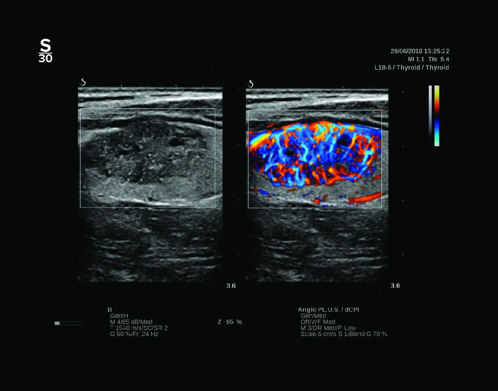
Angio PL.U.S.™

Angio PL.U.S.™

Planewave Ultrasensitive® Color Mode Imaging

  • Enhanced visualization and clarity in microvasculature and low velocity flow states.
  • Significantly improved color sensitivity and spatial resolution while maintaining exceptional 2D imaging.
  • Leverages the combination of UltraFast® imaging and 3D wall filtering to create a leap in Doppler imaging performance.
  • Ability to detect microvascularization in different types of lesions which opens the door to added clinical information and diagnostic perspectives.
TRIVU™

TRIVU™

Three answers in one.

Improve diagnostic confidence and enhance workflow efficiency with unparalleled insights into anatomy, stiffness, and blood flow in a glance.

This cutting edge, real time, simultaneous imaging mode displays:

  • B-mode
  • ShearWave® Elastography
  • Angio PL.U.S.™
UltraFast® Doppler

UltraFast® Doppler

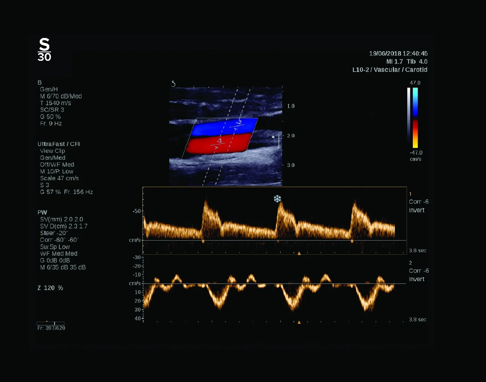
Uniting Doppler Technologies

SuperSonic® MACH®’s UltraFast® imaging has been leveraged to re-invent traditional modes of imaging by uniting ColorFlow Doppler with Pulsed Wave Doppler into a new mode called UltraFast® Doppler, expanding performance and capabilities.

  • Up to 300 frames per second in color Doppler to visualized transient phenomena missed by conventional techniques
  • Improved workflow with the ability to acquire multiple PW samples on the same acquisition
  • Superior post-processing capabilities
  • No limitation of color box size
  • Improved diagnostic confidence
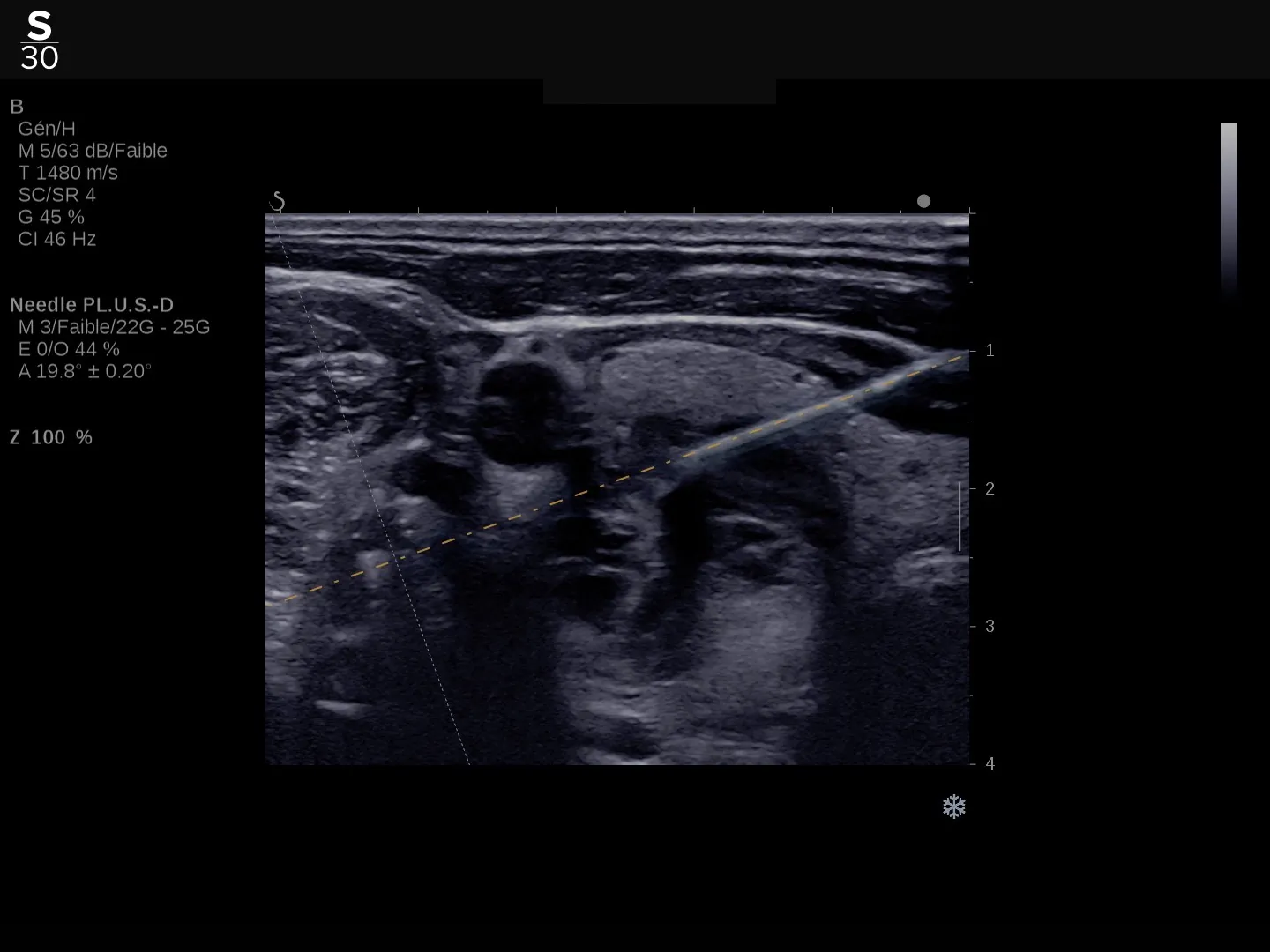
Needle PL.U.S.™

Needle PL.U.S.™

Needle Enhancement Imaging and Path Prediction

  • Enhanced Visualization: Better real-time visibility of biopsy needles without compromising B-mode image quality.
  • Trajectory Prediction: Unmatched precision in predicting needle paths, even with conventional needles.
  • Increased Confidence: Boosts procedural confidence for clinicians.
Liver Assessment Tools

Liver Assessment Tools

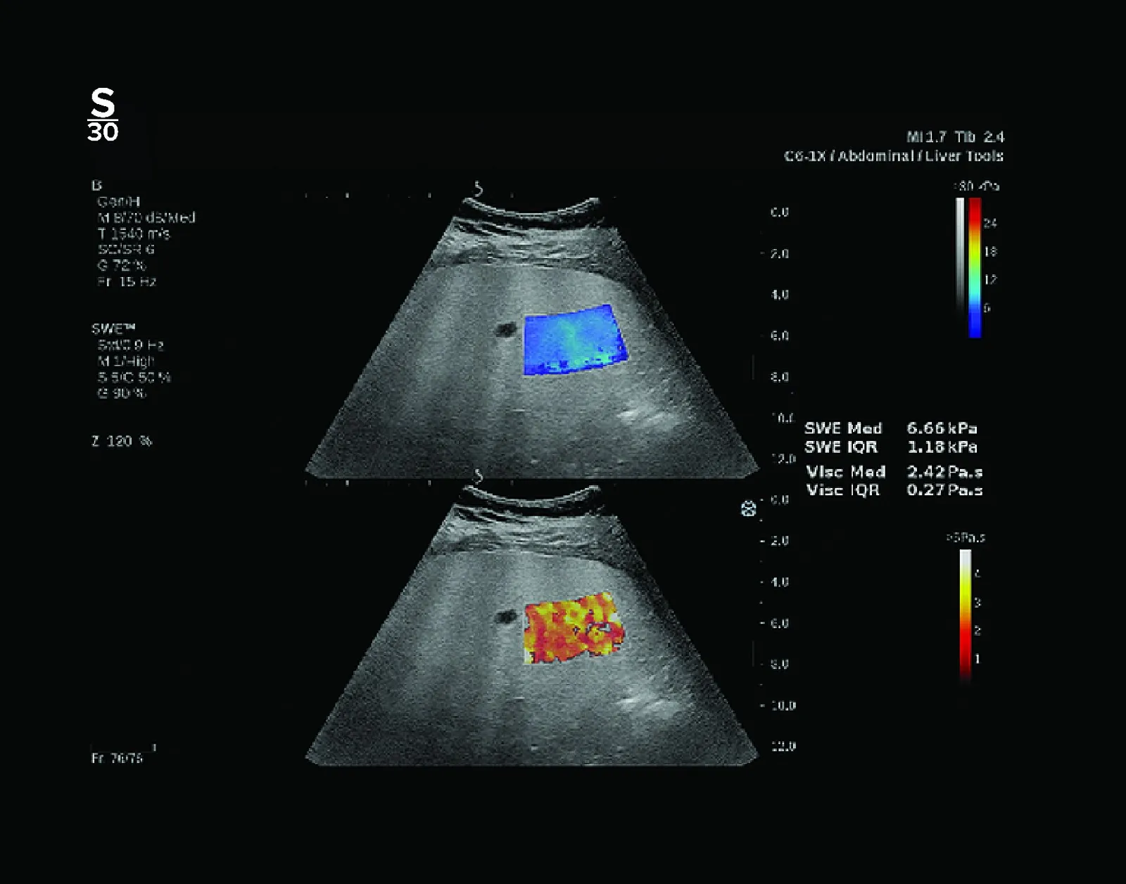
Liver Disease Assessment Suite

  • Quantitative tools for evaluating chronic liver disease in a non – invasive manner
  • ShearWave® PL.U.S.™ Display and quantification in real-time of liver fibrosis
  • Att PL.U.S.™ Quantification of beam attenuation, to stage steatosis
  • Ssp PL.U.S.™ Measurement of tissue sound speed related to fat content
  • Vi PL.U.S.™ Visualization and quantification of tissue viscosity. Displays ShearWave® dispersion, potential for liver inflammation
  • Full exam captured within 60 seconds.

Ultrasound Matrix Imaging

Enhanced real-time ultrasound biomarkers and image clarity for superior clinical value

  • Enabling new ultrasound biomarkers
  • Next generation image quality
  • Crisper, more detailed ultrasound images
  • Informed diagnostic decision support
  • Precise quantification of ultrasound data
  • Enhanced procedural guidance and accuracy
SonicPad™

SonicPad™

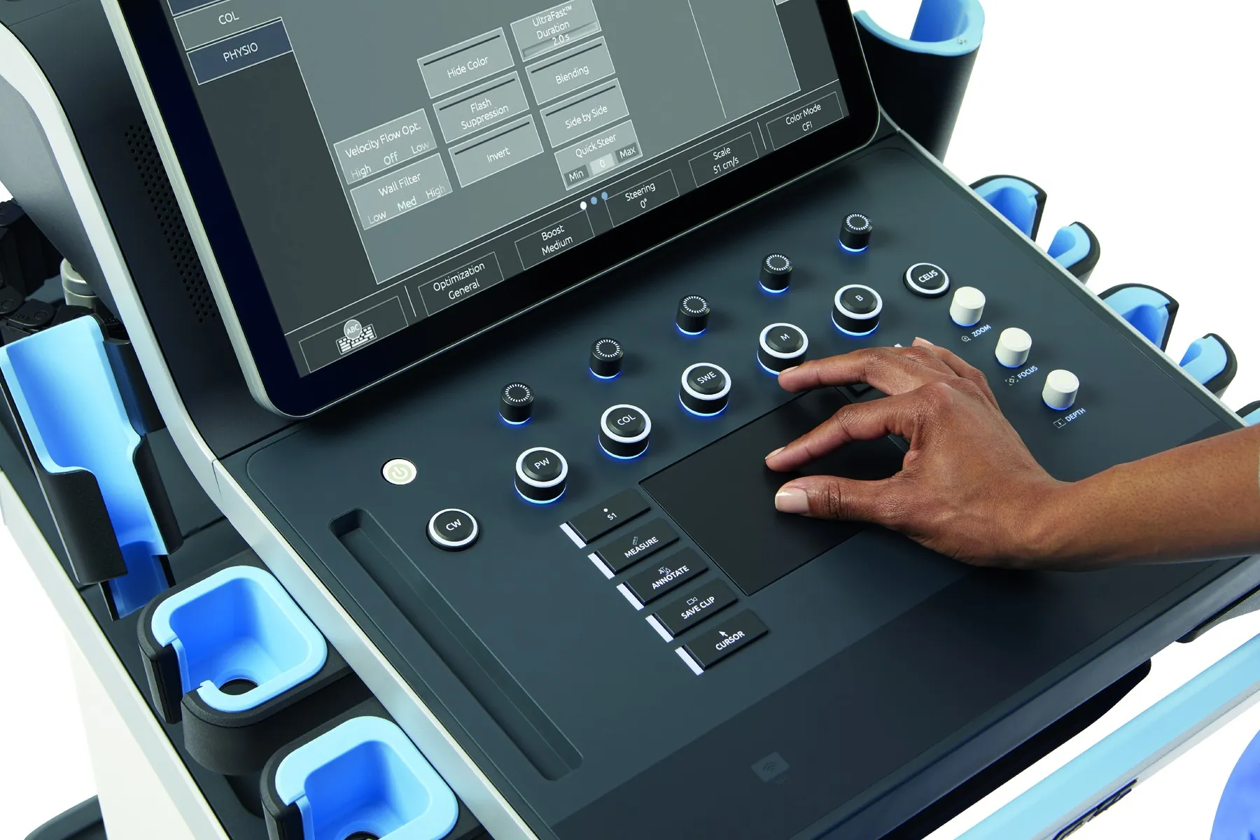
A new interface you've already mastered.

Minimize actions and movements for increased workflow and ergonomics with over 25 programmable gestures.

  • Our modern, embedded touchpad uses a familiar interface like the kind you already use on your smartphone, tablet or laptop.
  • Focus more on what you see and less on wrist fatigue or cleaning a trackball.

Click

The touch pad click function allows users to enables rapid access to key functions with a single touch.

Box move

Box move feature allows users to easily reposition and resize diagnostic boxes.

Sample Volume Size

The touch SV size feature allows users to adjust the sample volume size.

Clip Navigation

The clip navigation feature enables quick and easy navigation during multiple functions.

Zoom in

The stretch gesture allows users to magnify specific areas of an image.

Zoom out

The pinch gesture allows users to reduce the magnification of an image.

Ergonomics

Ergonomics

System Features

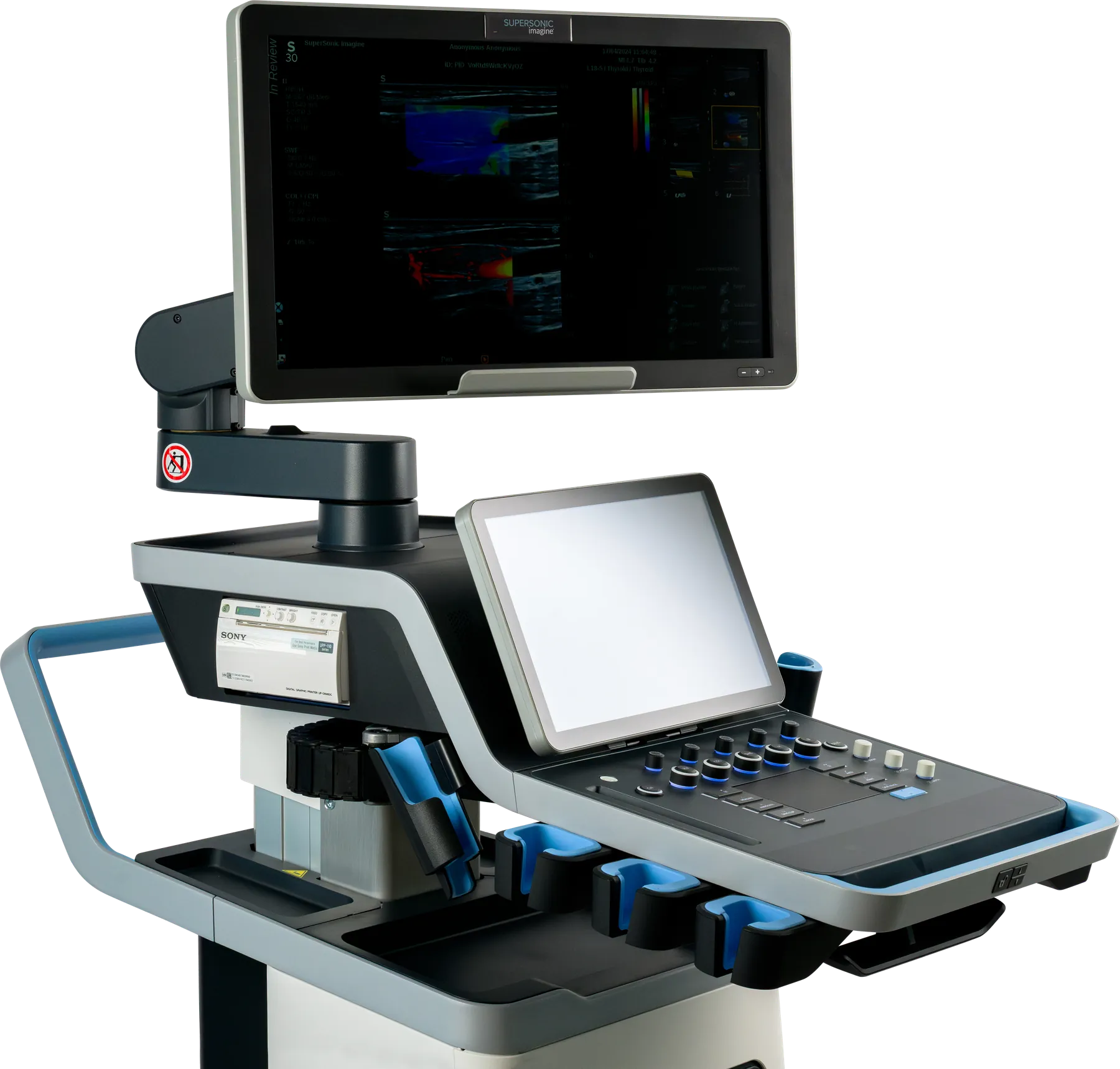
  • Streamlined console
  • Customizable buttons and knobs
  • SonicPad™
  • Low-level noise output
  • Lightweight transducers
  • 15" Full HD adjustable touchscreen
  • 23" Widescreen Full HD Monitor
  • Three-position central break
  • Large back handle for maneuvering
  • Pinless Connector Ports
  • Integrated gel warmer with auto-management of heat

Exam Optimization

Reduce Clicks

Users estimate 44% fewer clicks per ultrasound exam when using the SuperSonic® MACH® Ultrasound System’s Automated Protocols feature.

Reduce Time

On average the SuperSonic® MACH® Ultrasound System’s Automated Protocols feature reduced the time to perform an abdominal exam from 25 minutes to 11 minutes.

  • 100%of users strongly agree or agree that the SuperSonic® MACH® Ultrasound System’s Automated Protocol feature increases exam efficiency and image acquisition efficiency.
  • 100%of users strongly agree or agree that the SuperSonic® MACH® Ultrasound System’s Automated Protocols will prevent them from missing an image or step in the workflow, therefore providing radiologists more confidence.

SuperSonic Imagine® Data on File. SuperSonic® MACH 40 Ultrasound System CLT Claims Study. MISC-08417 Rev 001, 2022.

Query & Retrieve

Retrieving prior imaging can be very helpful when used for reference or comparison to the same area of interest in an ultrasound image.

  • Query the PACS server for previous exam(s)
  • Side-by-side on-cart display
  • Any retrieved exam can be displayed along with a live image in a current exam
  • Supported modalities: Ultrasound, Mammography, CT, MRI, Xray, Angiography, Nuclear Medicine and PET
  • Fly-through large 3D data sets acquired as series of 2D slices
  • Automated query and retrieve linked to MWL and use filters for specific data types
  • User may also push studies from PACS workstation to your SuperSonic® MACH® system

Reporting

Dedicated measurements and reporting tools

  • Fully customizable report
  • Dedicated worksheets for Liver, Breast, Vascular, OB-GYN
  • Selection of publications with cut off value per etiology
  • BI-RADS®
  • TI-RADS
  • 97%of users strongly agree or agree that SuperSonic® MACH® Ultrasound System’s report feature and worksheet includes all information needed to replace handwritten and computer-generated worksheets
  • 97%of users strongly agree or agree that SuperSonic® MACH® Ultrasound System’s report allows for a greater ability to customize images, measurements, patient history, and results in the final report.
  • 91%of users strongly agree or agree that it has never been easier to de-identify and anonymize a patient’s exam than when using the SuperSonic® MACH® Ultrasound System’s “Hide Patient” feature.

SSI Data on File. SuperSonic® MACH® 40 Ultrasound System CLT CLaims Study. MISC-08417 Rev 001, 2022.