TECHNOLOGY
SuperSonic Imagine holds 30 international patent families protecting its unique ultrasound imaging technology around the world.
See the list of patents
- UltraFast Imaging
The SuperSonic™ MACH™ range is equipped with UltraFast technology wich allows for the acquisition up to 20,000 frames per second1, this technology offers new possibilities for patient management. The next generation ofthe UltraFast technology — has 5x more computing power2. This unique technology is the foundation for several innovations which have changed the paradigm of ultrasound imaging: Real-time ShearWave™ Elastography (SWE), UltraFast Doppler, Angio PLUS – PLanewave UltraSensitive™ imaging and TriVu: real-time simultaneous B, SWE and Color+ imaging.
Clinical advantages of the UltraFast Imaging
- ShearWave Elastography
Real-time ShearWave Elastography, pioneered by SuperSonic Imagine, allows physicians to visualize and quantify the stiffness of tissue in a real-time, reliable, and reproducible manner. Tissue stiffness has become an important parameter in diagnosing potentially malignant tissue or other diseased tissue. Over 600 peer-reviewed publications have demonstrated the value of SWE for the clinical management of patients in a wide range of diseases.
- Angio PLUS - PLanewave UltraSensitive™ imaging
Angio PLUS provides a new level of microvascular imaging through significantly improved color sensitivity and spatial resolution while maintaining exceptional 2D imaging. Angio PLUS leverages the combination of ultrafast imaging and 3D wall filtering to create a leap in ultrasound Doppler imaging performance. Thanks to its ability to detect microvascularization in different types of lesions this new mode could open the door to added clinical information and diagnostic perspectives, in both benign and malignant lesions.
- TriVu
TriVu is a cutting edge real-time simultaneous imaging mode never seen before. TriVu combines B-mode, SWE and Color+ imaging, allowing physicians to visualize anatomy, tissue stiffness, and blood flow in the breast tissue concurrently. This unique mode also allows clinicians access to vascularization-guided measurement of tissue stiffness.
- Needle PLUS
Needle PLUS enables you to visualize both biopsy needles and anatomical structures in real time with unrivaled precision, and also predict where the needle is supposed to go. You save time, gain in comfort and reliability.
- UltraFast Doppler
UltraFast Doppler combines Color Flow Imaging and Pulsed Wave Doppler into one simple exam, providing physicians with exam results simultaneously and helping to increase patient throughput.
- Ultrasound Liver Markers
Three new quantitative tools that will open new perspectives in evaluating chronic liver disease in a non-invasive manner3,4,5: Att PLUS and SSp PLUS for evaluating ultrasound beam attenuation in the liver and Vi PLUS for visualization and quantification tissue viscosity.
- 3D Ultrasound Imaging
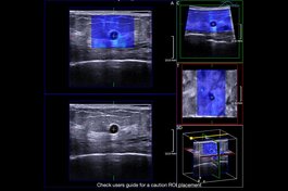
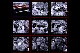
The SuperSonic MACH ultrasound systems now provide access to high-resolution B-mode and ShearWave PLUS elastography 3D acquisitions. 3D ultrasound imaging opens the door as an additional application in breast diagnostics and may support accurate interpretation.

|

|
| MultiPlanar display | MultiSlice display |
References:
1 Bercoff J, Ultrafast Ultrasound Imaging. Ultrasound Imaging - Medical Applications. 2011 Aug; DOI: 10.5772/19729
2 In comparison with Aixplorer® MultiWave™ systems
3 Fujiwara et al., The B-mode image-guided ultrasound attenuation parameter accurately detects hepatic steatosis in chronic liver disease, Ultrasound in Med. & Biol. 2018
4 Dioguardi Burgio et al., Ultrasonic Adaptive Sound Speed Estimation for the Diagnosis and Quantification of Hepatic Steatosis: A Pilot Study, Ultraschal Med. 2018
5 Deffieux T et al., Shear Wave Spectroscopy for In Vivo Quantification of Human Soft Tissues Visco-Elasticity, IEEE Transactions on Medical Imaging, 2009


