ABDOMEN
|
|
|
|
|
|

|
Consistency Matters
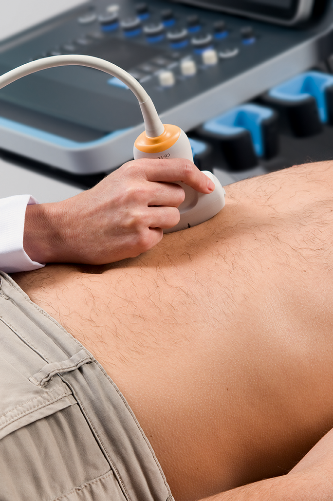
The SuperSonic™ MACH™ ultrasound systems bring you dedicated solutions for your abdominal and pelvic imaging challenges.
In general radiology, ¾ of all ultrasound exams are performed in the abdominal region. What if you could consistently identify abnormalities for a multitude of exams?
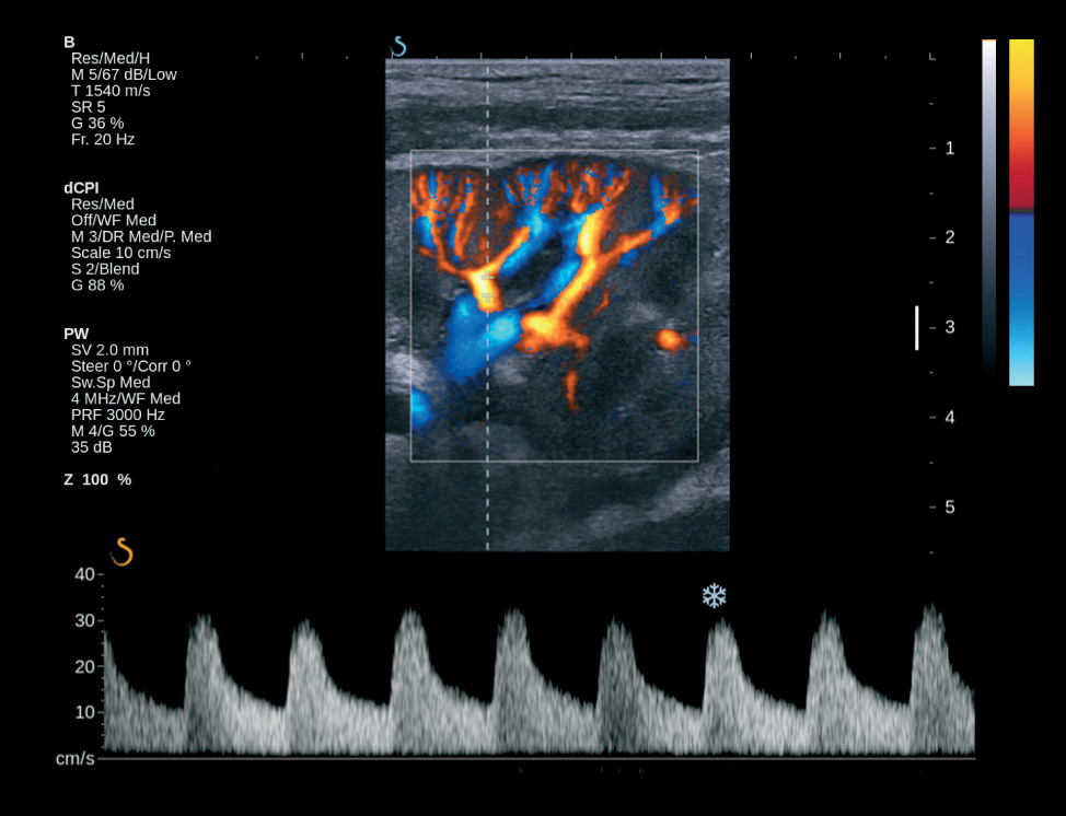
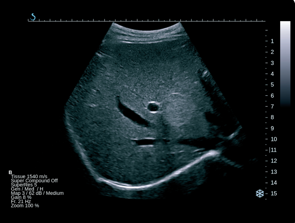
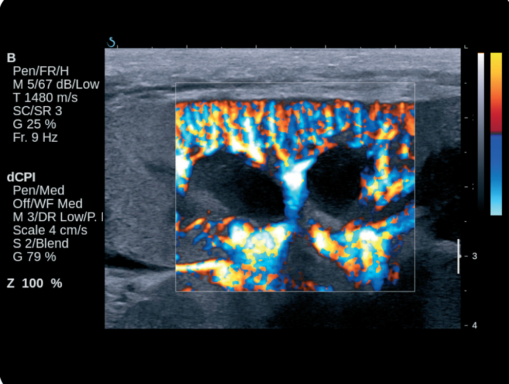
Unprecedented image quality is available with tools such as Tissue Harmonic Imaging to reduce speckle and enhance borders or Color Doppler with ultra sensitive flow in both superficial and deep abdominal structures. Additionally, flash suppression renders enhanced vascularization with no compromises in B-mode.
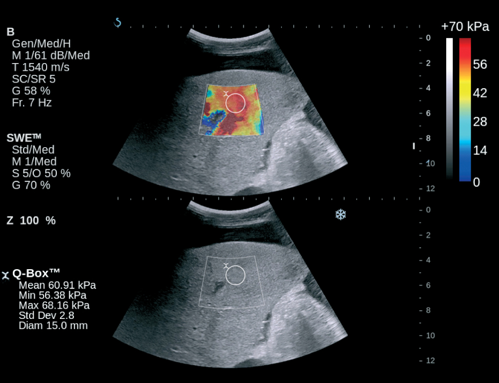
Real-time, patented ShearWave Elastography™ (SWE) gives you a diagnostic advantage with non-invasive, tissue elasticity measurements of abdominal organs such as the liver. For the first time ever, a 2D quantifiable image of tissue stiffness can be assessed in patients with suspected chronic or focal liver disease.
- A SWE map of liver elasticity shows lesion heterogeneity to help you accurately determine stiffest areas for measurement and biopsy. This color-coded map can help to avoid performing incorrect measurements in the kidneys or in near-by vessels
- Liver elasticity measurements can be obtained in challenging cases of obesity and ascites.
- ShearWave Elastography can also track changes in stiffness of liver or kidneys post treatment
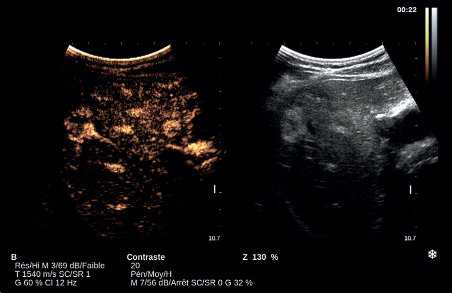
Contrast Imaging
SuperSonic MACH systems also provide advanced and comprehensive Contrast Enhanced Ultrasound (CEUS) solutions for detection, characterization, and monitoring of solid tumors particularly in the Liver and Abdomen. Aixplorer MACH are the only system, which can do both CEUS and ShearWave Elastography to enhance the comparison of blood flow in the microcirculation with the mechanical and structural properties of tissue, giving more diagnostic information.
Long loop capture that guarantees up to five minutes of contrast acquisition time in both prospective and retrospective captures.
Flash imaging, contrast timer, MVI and POI also available.
Transducers

| Single Crystal Curved Array XC6-1
Number of elements: 192 | ||

| SuperLinear™ SL18-5
Number of elements: 256 | ||

|
SuperLinear™ SL10-2
Number of elements: 192 | ||

| SuperLinear™ SL15-4:
Number of elements: 256 | ||

| Single Crystal Phased Array XP5-1
Number of elements: 96
|

