BREAST
|

|
|
|
|
|
|
|
|

|
Comprehensive Imaging Solution for the Management of Breast Cancer Patients
SuperSonic Imagine has raised the bar in breast imaging.
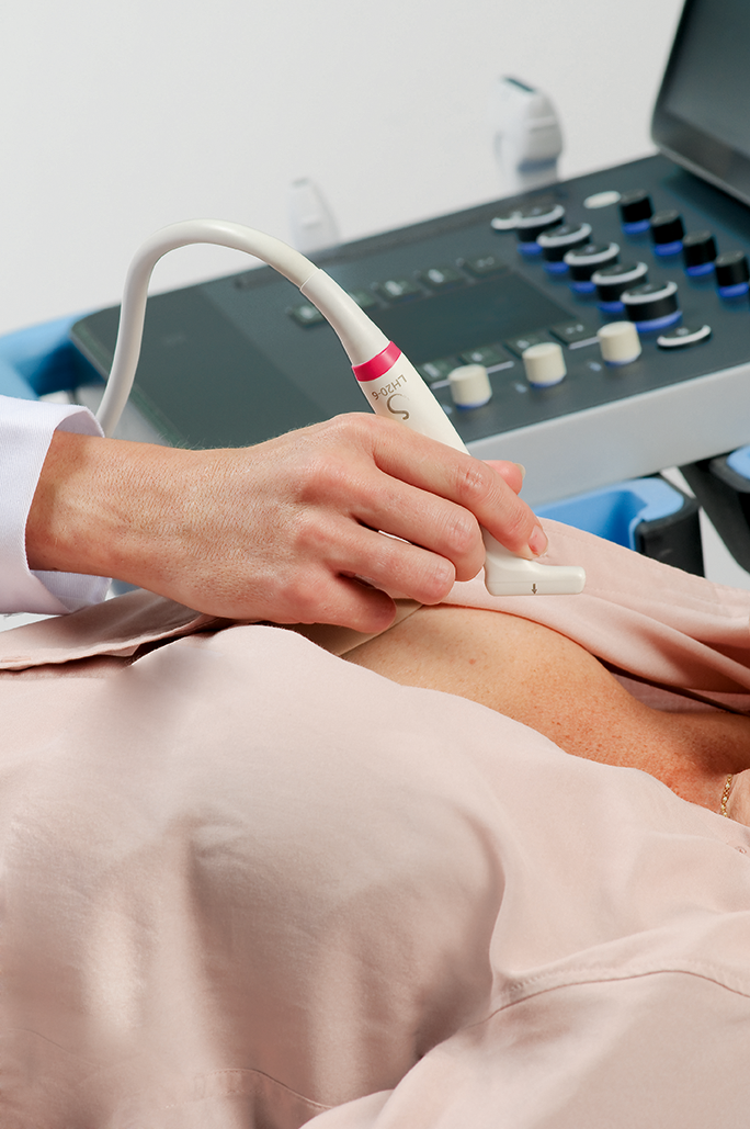
With SuperSonic MACH ultrasound systems, breast specialists now have access to:
- Premium image quality through a comprehensive suite of breast optimized transducers
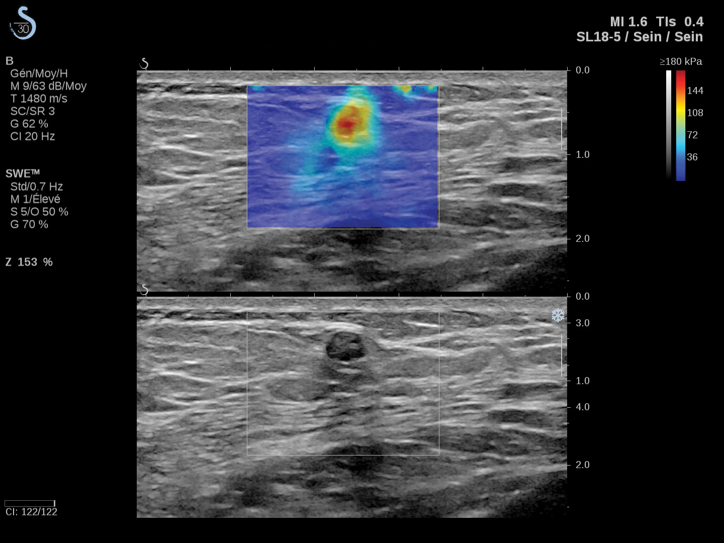
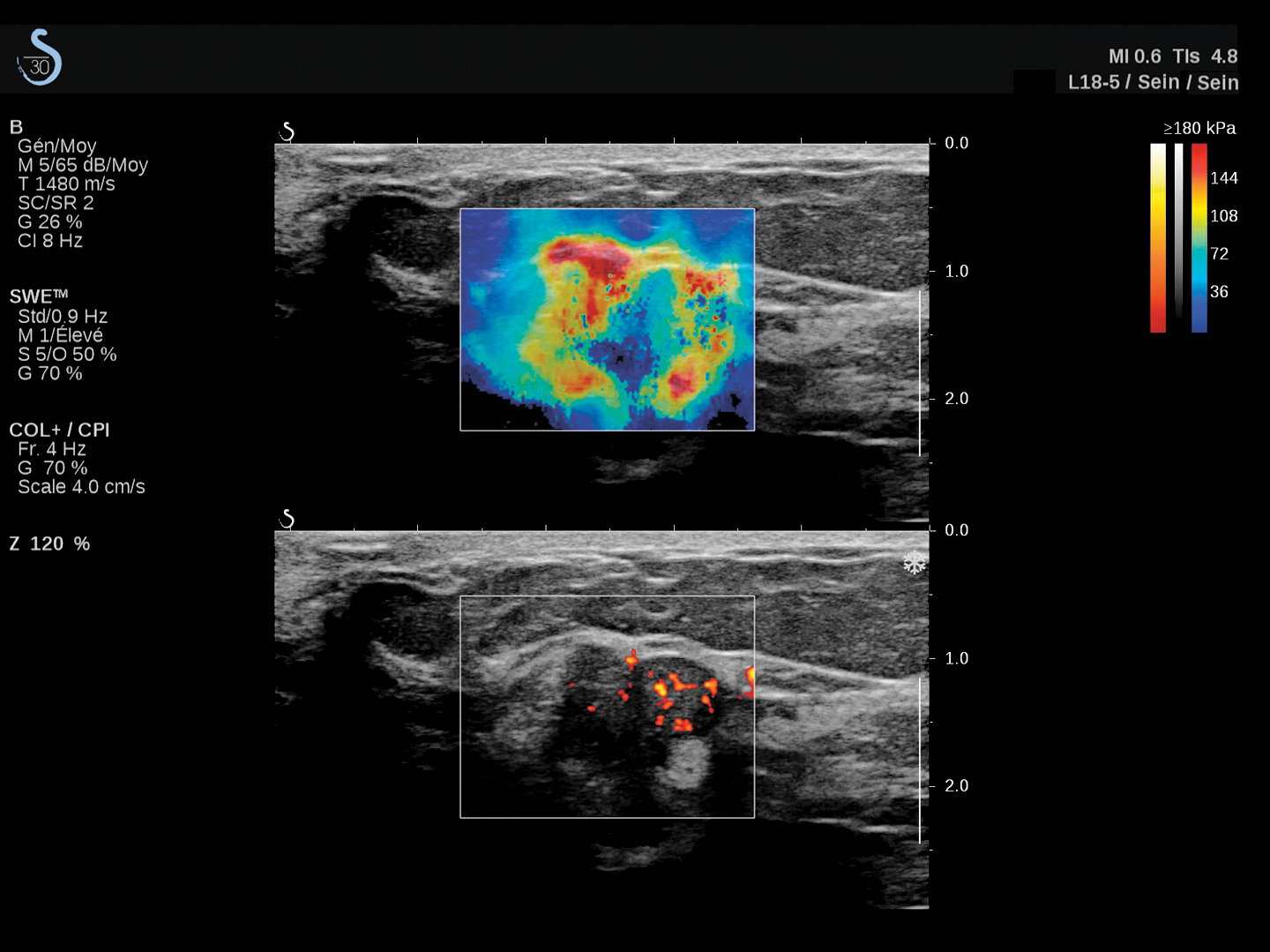
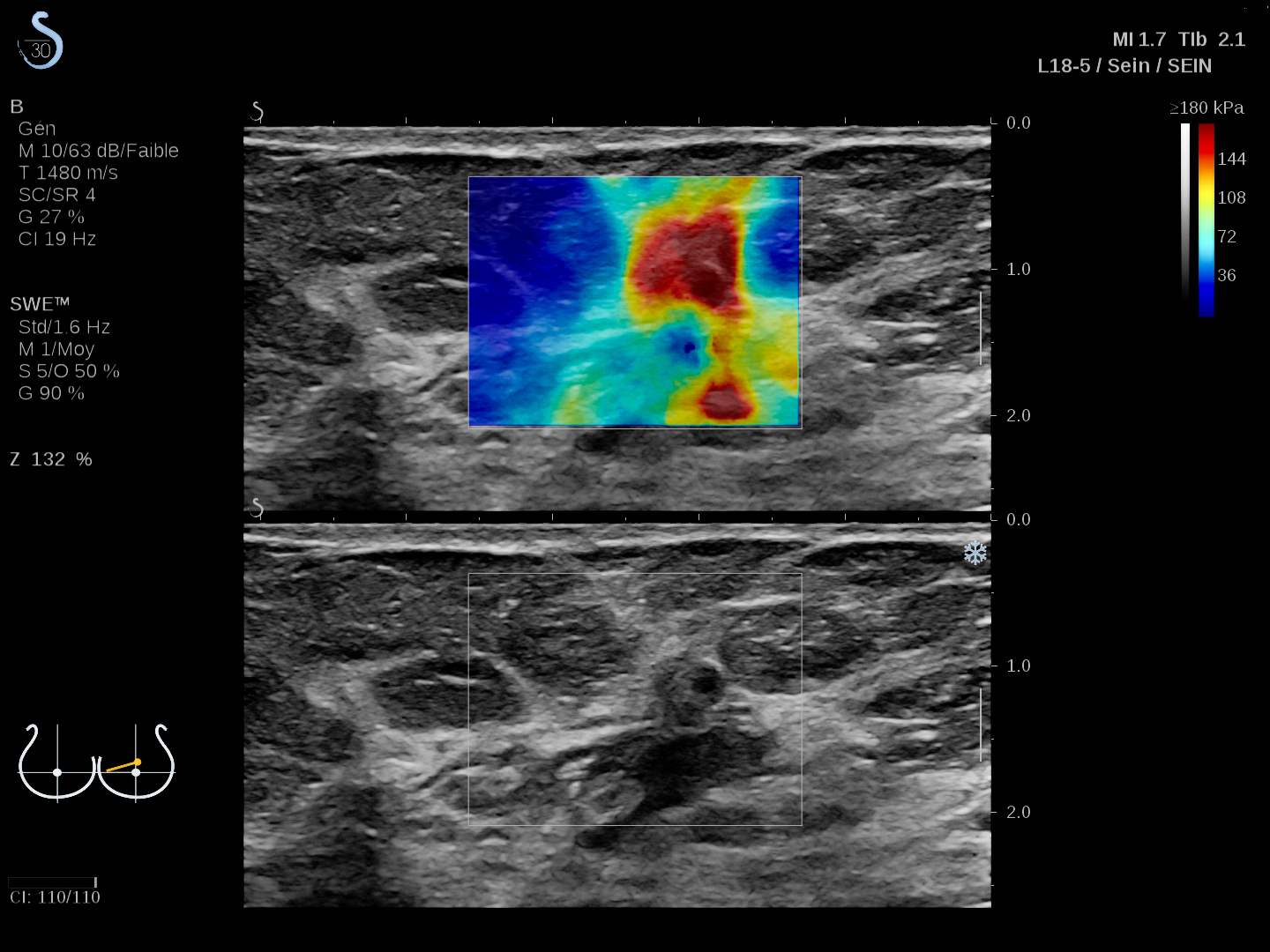
- Real-time ShearWave™ Elastography, the most clinically studied shear wave-based elastography technology for breast lesion characterization
- 3D breast software-based application that offers unique visualizations of breast anatomy and detailed characterization of lesions
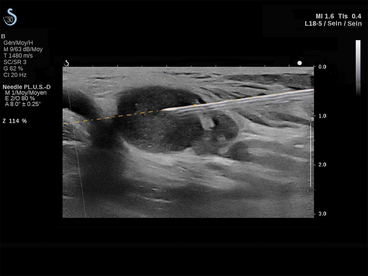
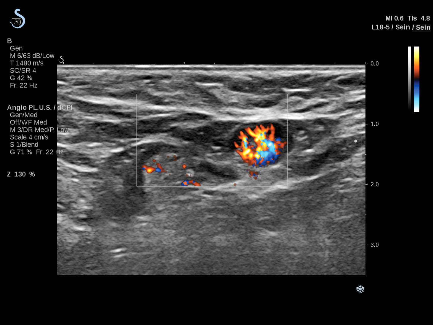
- Innovations including: Angio PLUS, Trivu and Needle PLUS
ShearWave™ Elastography (SWE) has been proven to be a complementary tool for the management of breast cancer patients for:
- lesion characterization1
- surgical planning2
- therapy monitoring3
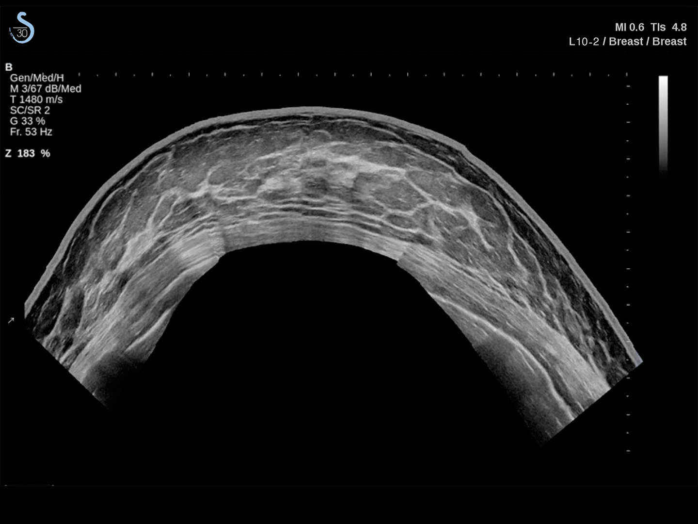
3D Ultrasound Imaging is designed to enhance diagnostic certainty. Lesion characteristics and morphometry can be leveraged together with the coronal plane imaging to provide supplementary information.
- May provide greater diagnostic information due to whole volume images and the unique coronal plane.
- Resolution is maintained at all depths. For example, lesions next to the pectoralis muscle are easily visible to the clinician.
- It may also assist physicians in the workup of difficult lesions, such as patients with dense breasts.4

|

|
| MultiPlanar display | MultiSlice display |
Transducers

| SuperLinear™ SL18-5
Number of elements: 256
| ||

| SuperLinear™ SLH20-6
Number of elements: 192 | ||

| SuperLinear™ SL10-2
Number of elements: 192 | ||

| SuperLinear™ SL15-4
Number of elements: 256 | ||

| SuperLinear™ Volumetric SLV16-5
Number of elements: 192 |
References:1 Berg WA et al. Radiology. 2012 Feb;262(2):435-49 2 Mullen R et al. Clin Radiol. 2014 Dec;69(12):1259-63 3 Lee SH et al. Ann Surg Oncol. 2015 Dec;22 Suppl 3:376-84 4 Berg WA, Blume JD, Cormack JB, et al. Combined screening with ultrasound and mammography vs. mammography alone in women at elevated risk of breast cancer [published correction appears in JAMA. 2010 Apr 21;303(15):1482]. JAMA. 2008;299(18):2151-2163. doi:10.1001/jama.299.18.2151

